Clinical utility of BAL
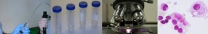
Alterations in BAL fluid and cells reflect pathological changes in the lung parenchyma. BAL has become a widely applied diagnostic tool in pulmonary medicine. The BAL procedure was developed as a research tool. Meanwhile its usefulness, also for clinical applications, has been appreciated worldwide in diagnostic work-up of infectious and non-infectious infiltrative and immunological lung diseases. Moreover, BAL has several advantages over biopsy procedures. It is a safe, easily performed, minimally invasive, and well tolerated procedure. In this respect, when the clinician decides that a BAL might be helpful to provide diagnostic material, it is mandatory to consider the provided information obtained from BAL fluid analysis carefully and to have reliable diagnostic criteria. Therefore, knowledge how to interpret BAL fluid cytology is mandatory to improve the diagnostic power. Additionally, BAL can play a very important role in the diagnosis of respiratory infections, and it is useful in monitoring the lung allograft.
BAL Guidelines
Barriers which tried to restrict the use of BAL to research application and to put down its clinical value have finally been overcome. In two published international statements (ATS, ERS, also WASOG) on the major interstitial lung diseases, BAL was considered to be helpful in strengthening the diagnosis in a sarcoidosis patient without biopsy; BAL and/or transbronchial biopsy were considered as a requirement to exclude other diseases in a patient with idiopathic pulmonary fibrosis/UIP who does not undergo surgical biopsy (one of the four major criteria for making a clinical diagnosis of the disease).
Interpretation of BAL fluid analyses results
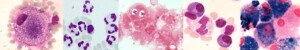
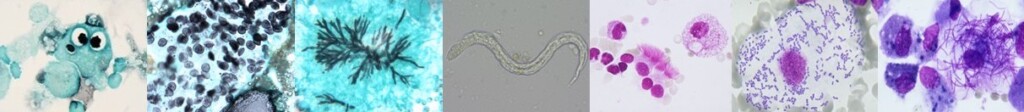
BAL studies should not be limited to counting the cell differentials only. At least as important as looking at cell differentials is to observe the morphological appearances of cells and particles. Examination of BAL cells or acellular components of BAL fluid via gene microarray technology or proteomic analyses may allow BAL to assume a more prominent role in diagnosis and management of lung disease in the near future. In the follow-up depicting prognosis and response to treatment BAL fluid analysis has less clinical relevance. Examples are the different morphology in extrinsic allergic alveolitis (foamy macrophages, heterogeneous macrophage size, presence of plasma cells) versus that of sarcoidosis (more monomorphous appearance of macrophages, less activated lymphocytes), the presence of malignant cells, the characteristic features of alveolar proteinosis, or dust particles such as asbestos bodies, and other features.
Assess BAL fluid analyses results always in the context of all clinical data
Also, it is important to consider BAL cell differentials not in isolation but in the context of the clinical setting and the radiological, particularly the HR-CT appearance of the disease. For example, if the CT scan shows a patchy ground glass pattern, BAL may be able to reveal that this patient suffers from extrinsic allergic alveolitis (high lymphocyte count), or a smoking related respiratory bronchiolitis/interstitial lung disease (high smoker’s macrophage count and normal cell differential), or alveolar haemorrhage (high count of haemosiderin laden macrophages). Although details hidden in BAL fluid may add useful information about a patient’s disorder, the results should be considered in the context of other information from conventional investigative methods and the individual’s unique history. To establish the diagnosis a thorough history is essential as it may identify a potential aetiological factor.
© ild care foundation
This course addresses various topics.
By means of web lectures, case studies, and reading papers you will learn:
- how to use guidelines to facilitate appropriate interpretation of cellular BALF analysis results
- how to use information regarding the clinical utility of cellular BALF analysis to establish or ruling out a certain diagnosis
- how to recognize more or less disease specific features and limitations of the BAL
KEY NOTES
- BAL fluid cellular profile analysis is useful in the diagnostic work-up of diffuse lung damage
- BAL fluid analysis is helpful in excluding or diagnosing infectious disorders
- BAL fluid analysis should not be limited to counting the cell differentials only
- At least as important is to observe the morphological appearances of cells and particles
- BAL fluid analysis may avoid more invasive diagnostic procedures in certain cases
- To establish the correct diagnosis a thorough history is essential as it may identify a potential aetiological factor
- BAL cell differentials should always be considered in the context of clinical information and the radiological, particularly the HR-CT appearance of the disease
Videos
Video 1. Role of Bronchoalveolar Lavage (BAL) in the diagnostic work-up of patients with interstitial lung diseases (ILD)
Video 1
Download PDF of Video 1
Video 2. Work-up of BAL fluid in the laboratory
Video 2
Download PDF of Video 2
3. Introduction diagnostic value of cellular analysis of BAL fluid
Video 3
Download PDF of Video 3
4. Diagnostic value of BAL fluid analysis in infectious diseases
Video 4.
Download PDF of Video 4.
5. Diagnostic value of BAL in sarcoidosis
Video 5.
Download PDF of Video 5.
6. Diagnostic value of BAL in EAA
Video 6.
Download PDF of Video 6.
Learning Cases
Case report I
Case report II
Case report III
Case report IV
Case report V
Case report VI
Case report VII
Case report VIII
Recommended readings
References Nederlands
Drent M, Costabel U. BAL in de diagnostiek van diffuse longaandoeningen. Ned Tijdschr Geneeskd 1998; 142(49):2661-5.
Figures Kitty Linssen ild care today 2009 (3)
References English
Publications on BAL Kitty Linssen
Publication on BAL Marjolein Drent
Marjolein Drent (1955) studied physical therapy at the Arnhem Academy of Physical Therapy, the Netherlands(NL); she graduated in 1979. She finished her medical study in 1988 at the Catholic University of Nijmegen, NL. In 1993 she completed her thesis entitled: ‘Diagnostic value of bronchoalveolar lavage in interstitial lung diseases (ILD).’ After she finished her training as a pulmonologist in 1994 she was appointed as a pulmonologist till she retired in July 2022. Currently, she is professor emeritus of ILD at the Department of Toxicology of the Faculty of Health and Life Sciences of the University of Maastricht, NL. She is the founder and chair of the ild care foundation (please visit: www.ildcare.nl)
Contact: info@ildcare.nl

In Memoriam: Catharina (Kitty) Linssen (1974–2025)
Catharina (Kitty) Linssen was born on May 19, 1974, in Roermond, the Netherlands. She studied Medicine at Maastricht University, obtaining her medical degree in August 2000. In the same year, she began her professional journey as an AGNIO at the Department of Medical Microbiology of Maastricht University Medical Center (MUMC), Maastricht, NL. A year later, she commenced her specialist training in Medical Microbiology at the same department.
During her training, Kitty undertook a research project on the Diagnostic Value of Bronchoalveolar Lavage in Infectious Disorders, combining conventional diagnostic methods (microscopy, culture, and serology) with innovative molecular techniques.
She also created stunning images of cells and infectious agents, combining scientific precision with remarkable visual artistry (please read also: https://ildcare.nl/Downloads/ildcaretoday/ildcaretoday2009_3_Linssen.pdf).
Her dedication and scientific curiosity culminated in the successful defense of her PhD thesis in 2007.‘Diagnostic value of bronchoalveolar lavage in infectious disorders’.
Until January 2012, Dr. Linssen contributed as a staff member at the Department of Medical Microbiology of MUMC. From February 2012 until July 2025, she was a respected and highly valued member of the Department of Medical Microbiology at Zuyderland Medical Centre, Heerlen, NL, where her expertise, collegiality, and warm personality left a lasting impact on colleagues and patients alike.
On July 27, 2025, Kitty passed away. She will be remembered not only for her significant scientific contributions but also for her kindness, dedication, and the positive influence she had on everyone around her. Her legacy continues to inspire those who knew her.
© ild care foundation